Capturing details of brain cells on a nanometer scale, the researchers found evidence that the neurons of people with schizophrenia could have unique differences in thickness and curvature, and this could even explain some of their symptoms.
The finding comes from an analysis on just a small handful of donors and is a long way from demonstrating how contrasting nerve cell structures could explain the neurological condition.
But as we understand these unusual features grow, it could lead to better treatment methods, helping to provide tens of millions around the world with a better quality of life.
The study, led by researchers at Tokai University in Japan, used two different X-ray microscope technologies, one at the SPring-8 light source in Japan, the other at the Department of Energy’s Advanced Photon Source (APS). US.
Both accelerate the particles along curved paths in what is known as the synchrotron, causing them to pour short wavelengths of electromagnetic radiation into the X-ray part of the spectrum.
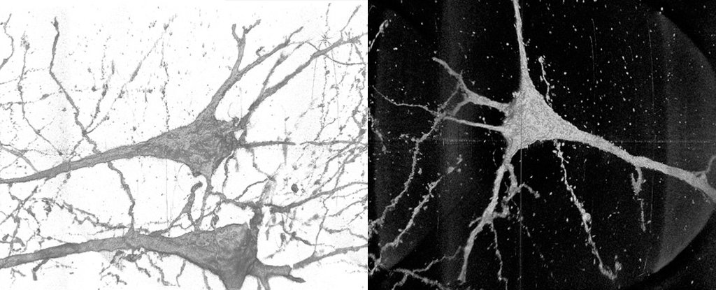
Using X-rays as a source of radiation to photograph the fine details of small objects – such as neurons – can be a double-edged sword.
On the one hand, their tight wavelengths are just what each buckle and tissue of a cell membrane captures. APS is capable of a resolution down to 10 nanometers, a scale that brings it remarkably close to revealing the texture of the individual protein channels that pepper a cell membrane.
Viewed from sufficient angles, it is possible to reconstruct neurons as high-definition three-dimensional terrain.
Unfortunately, no matter how small the neurons are, they are also quite long. Tracking every swelling of their surface is a tedious task when you have to sneak along the entire millimeters of their body.
“The sample must travel through the X-ray beam to track the neurons through the sample,” says Vincent De Andrade, a physicist in Argonne’s X-ray science division.
“The field of view of our X-ray microscope is about 50 microns, about the width of a human hair, and you have to follow these neurons for several millimeters.”
Taking tissue samples from a selected part of the brain in four deceased people diagnosed with schizophrenia and four without, the team undertook the tedious work of scanning nerve cells using the two different synchrotron facilities.
The images were combined to reconstruct neurons as digital models, which contributed to a larger data set that could be statistically compared and contrasted in search of distinctive features.
They found, statistically speaking, the thickness and curvature of cellular features extending away from the body of the neuron was significantly different among people with schizophrenia compared to those without the condition.
These variations could affect the way neurons transmit messages along their length, which could go a way to explain the characteristics of the disorder, which in its worst forms includes hallucinations, impaired motor control, and illusions.
Exactly what lies behind such deviations in cell geometry, or if the variations extend to the synaptic “toes” of the neuron, will require even more detail than the current generation’s synchrotron can handle.
This could change when APS obtains an update of 815 million US dollars over the next few years, which will see it produce X-ray beams 500 times brighter than those it currently emits.
“Updating APS will allow for better sensitivity and resolution for imaging, making the process of mapping neurons in the brain faster and more accurate,” says De Andrade.
“We would need resolutions of better than 10 nanometers to capture synaptic connections, which is the holy grail for a comprehensive mapping of neurons, and they should be achievable with the upgrade.”
Joining the mechanisms behind the development of schizophrenia is a complex process that will require advanced imaging and computing technology.
Gradually, we come to understand the multitude of genetic and environmental factors that see that the brain still changes in the womb and continues to change as the baby grows to adulthood.
If there are ways in which this can be detected and treated early, we could help limit, if not prevent, the worst feature that can put people at risk for serious mental illness.
This research was published in Translational psychiatry.